Introduction
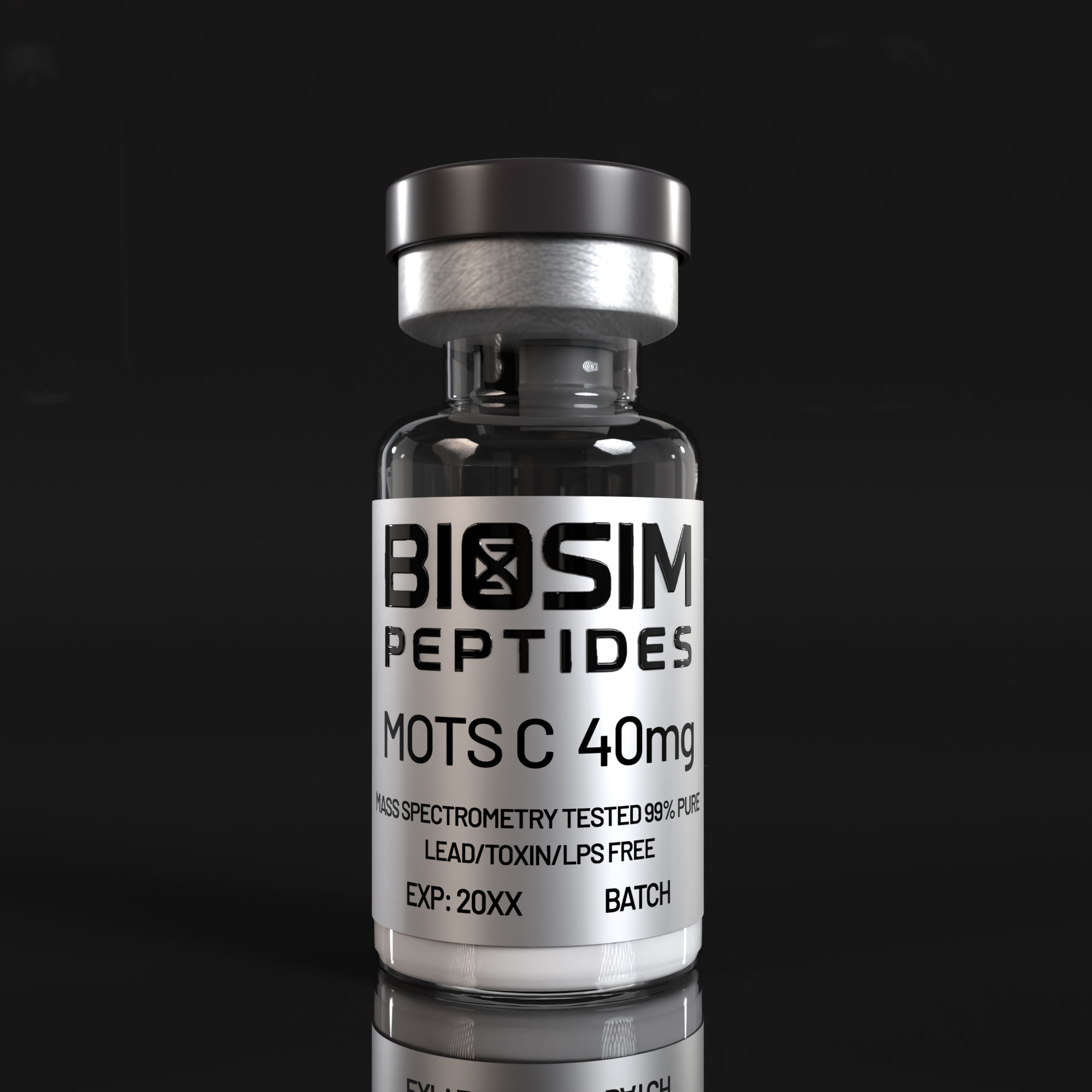
MOTS-c (Mitochondrial Open Reading Frame of the 12S rRNA-c) is a 16-amino acid peptide (Met-Arg-Trp-Gln-Glu-Met-Gly-Tyr-Ile-Phe-Tyr-Pro-Arg-Lys-Leu-Arg; MRWQEMGYIFYPRKLR) encoded within the 12S ribosomal RNA region of the mitochondrial genome. Its discovery in 2015 by Cohen and colleagues at the University of Southern California fundamentally expanded the paradigm of mitochondrial signaling: previously, the mitochondrial genome was understood to encode only 13 oxidative phosphorylation proteins, 2 rRNAs, and 22 tRNAs. The identification of MOTS-c — a biologically active peptide with systemic metabolic regulatory functions — established that mitochondria also encode signaling molecules capable of communicating organellar status to the nuclear genome, a process termed mitochondrial retrograde signaling [1][2].
MOTS-c belongs to a growing family of mitochondrial-derived peptides (MDPs) that also includes humanin, SHLP1-6, and small humanin-like peptides. Among these, MOTS-c is distinguished by its potent metabolic effects: it acts as an exercise mimetic, enhances insulin sensitivity, promotes glucose uptake via GLUT4 translocation, activates AMPK-dependent mitochondrial biogenesis, and suppresses systemic inflammation. Its mechanism of action involves stress-induced translocation from mitochondria to the nucleus, where it directly regulates nuclear gene expression — a feature that positions it at the nexus of mitonuclear crosstalk and metabolic adaptation [1][2].
Molecular Biology: Mitochondrial Retrograde Signaling
The mitochondrial genome of MOTS-c challenges the long-held view that mitochondria are solely ATP-producing organelles subordinate to nuclear control. Under conditions of metabolic stress — glucose deprivation, oxidative challenge, or exercise — MOTS-c translocates from the mitochondrial matrix to the nucleus via a mechanism that requires the mitochondrial permeability transition pore (mPTP) and importin-mediated nuclear localization. Once in the nucleus, MOTS-c binds to the antioxidant response element (ARE) in the promoter regions of stress-responsive genes and regulates their transcription. This represents a direct molecular conduit through which mitochondrial metabolic status is communicated to the nuclear epigenome without intermediary cytosolic signaling cascades [1][2].
The peptide directly interacts with the folate cycle enzyme methylenetetrahydrofolate dehydrogenase 2 (MTHFD2) and inhibits de novo purine biosynthesis, leading to accumulation of the purine synthesis intermediate AICA ribonucleotide (AICAR) — the endogenous activator of AMP-activated protein kinase (AMPK). This AMPK activation, in turn, phosphorylates and activates peroxisome proliferator-activated receptor gamma coactivator 1-alpha (PGC-1α), the master regulator of mitochondrial biogenesis. The resulting AMPK → PGC-1α → mitochondrial biogenesis axis explains how a mitochondrially encoded peptide feeds forward to enhance the very organelle that produced it, creating a homeostatic positive feedback loop [3].
MOTS-c Signaling Cascade
Metabolic stress → MOTS-c translocates from mitochondria to nucleus
→ Inhibits MTHFD2 → ↑ AICAR → AMPK activation
→ AMPK phosphorylates PGC-1α → mitochondrial biogenesis
→ AMPK promotes GLUT4 translocation → glucose uptake
→ Nuclear ARE binding → antioxidant gene expression
→ NLRP3 inflammasome suppression → anti-inflammatory
Metabolic Regulation: Exercise Mimetic Properties
MOTS-c has been characterized as an “exercise mimetic” based on its ability to recapitulate key metabolic adaptations normally induced by physical activity. Endogenous MOTS-c levels in skeletal muscle and plasma increase in response to acute exercise in both rodents and humans, and the peptide’s downstream effects — AMPK activation, GLUT4 translocation, enhanced fatty acid oxidation, and mitochondrial biogenesis — mirror the molecular signature of exercise training [1][3].
Glucose Homeostasis: MOTS-c promotes GLUT4 translocation to the plasma membrane in skeletal muscle cells through a mitofusin-dependent mechanism, enhancing insulin-independent glucose uptake. This effect has been demonstrated in both L6 myotubes and primary human skeletal muscle cells, with evidence that mitofusin-2 (MFN2) — a mitochondrial fusion protein — is required for the MOTS-c-induced GLUT4 trafficking [4].
Insulin Sensitivity: Clinical studies have characterized circulating MOTS-c levels across the metabolic health spectrum. MOTS-c concentrations are inversely correlated with insulin resistance (HOMA-IR), BMI, and inflammatory markers in human subjects, and are significantly reduced in individuals with obesity, type 2 diabetes, and polycystic ovary syndrome (PCOS). Importantly, the relationship appears bidirectional: lower MOTS-c levels predict metabolic dysfunction, and metabolic dysfunction further suppresses MOTS-c expression, creating a vicious cycle that may contribute to progressive metabolic decompensation [5][6].
Skeletal Muscle Protection and Performance
MOTS-c has emerged as a critical regulator of skeletal muscle health across diverse atrophy-inducing conditions. In immobilization-induced muscle atrophy models, MOTS-c administration significantly attenuated the loss of muscle mass and cross-sectional area by suppressing intramuscular lipid infiltration — a process that converts functional contractile tissue into non-contractile lipid-laden fibrotic tissue. The mechanism involves AMPK-mediated inhibition of lipogenic transcription factors (SREBP-1c, PPARγ) within the muscle microenvironment [7].
In glucocorticoid-induced atrophy — a clinically relevant model of steroid myopathy — both MOTS-c and the related MDP humanin attenuated dexamethasone-induced myotube atrophy in primary human skeletal muscle cells, with MOTS-c demonstrating superior potency at equimolar concentrations. The protective effect was mediated through suppression of the ubiquitin-proteasome system (MuRF1 and atrogin-1/MAFbx) and maintenance of mitochondrial membrane potential [8].
In cancer cachexia models (C26 adenocarcinoma), MOTS-c partially protected against skeletal muscle deterioration, preserving myofiber cross-sectional area and grip strength without affecting tumor burden. The selective protection of muscle tissue — without stimulating tumor growth — distinguishes MOTS-c from anabolic interventions like testosterone or GH that may inadvertently promote neoplastic proliferation [9].
Mitochondrial Bioenergetics: MOTS-c directly enhances intrinsic mitochondrial respiratory capacity. In skeletal muscle, MOTS-c treatment improves Complex I and Complex II-driven oxygen consumption, increases ATP synthesis rates, and enhances mitochondrial coupling efficiency (P/O ratio) in a PGC-1α/AMPK-dependent manner. These effects are observed even in the absence of changes in mitochondrial content, indicating that MOTS-c improves the functional quality of existing mitochondria in addition to promoting biogenesis [3].
Cardiometabolic and Anti-Inflammatory Effects
Beyond skeletal muscle, MOTS-c exerts protective effects on cardiac tissue. In diabetic rat models, MOTS-c suppressed both systemic and cardiac NLRP3 inflammasome activation, reducing circulating IL-1β and IL-18 levels and attenuating myocardial fibrosis. In hyperoxia-induced neonatal cardiac injury, MOTS-c inhibited oxeiptosis — a novel form of caspase-independent regulated cell death — by maintaining the KEAP1-PGAM5 interaction, preventing PGAM5-mediated dephosphorylation of the oxeiptosis executioner AIFM1 [10][11].
The anti-inflammatory effects of MOTS-c extend to systemic contexts. Reduced circulating MOTS-c levels have been documented in Hashimoto’s thyroiditis, with the degree of reduction correlating with both thyroid autoantibody titers and metabolic dysregulation markers, suggesting that MOTS-c deficiency may function as a biomarker linking autoimmune thyroid disease to its metabolic comorbidities [12].
Research Applications
Metabolic Disease Models: MOTS-c is an ideal probe for studying the mitonuclear communication axis in insulin resistance, obesity, and type 2 diabetes. Its endogenous regulation by metabolic state and its reciprocal control of systemic glucose homeostasis make it uniquely suited for investigations of the mitochondrial origins of metabolic disease.
Muscle Wasting and Sarcopenia: The consistent demonstration of MOTS-c’s protective effects across multiple atrophy models (immobilization, glucocorticoid, cancer cachexia) positions it as a lead candidate for mechanistic studies of muscle preservation. Unlike IGF-1/GH-based anabolic strategies, MOTS-c’s AMPK-mediated mechanism does not stimulate the PI3K-AKT-mTOR growth pathway, potentially offering tissue-specific protection without proliferative risk.
Exercise Physiology: As an endogenous exercise-responsive peptide with exercise-mimetic properties, MOTS-c is a valuable tool for dissecting the molecular pathways through which physical activity improves metabolic health — enabling studies that isolate specific exercise-responsive signaling nodes without the confounding variables of whole-body exercise interventions.
Cardiometabolic Research: MOTS-c’s dual cardioprotective and metabolic effects make it relevant to studies of diabetic cardiomyopathy, ischemia-reperfusion injury, and the cardiovascular complications of metabolic syndrome.
Safety and Handling
This product is supplied as a lyophilized powder for research purposes only. It is not for human consumption or diagnostic use. MOTS-c is a small, cationic peptide (pI ~11.2, MW 2,174 Da) that is soluble in water and physiological buffers. Store lyophilized product at -20°C in a desiccated, light-protected environment. Reconstitute with sterile water, phosphate-buffered saline, or appropriate buffer at neutral pH. Due to the peptide’s small size and absence of disulfide bonds, MOTS-c is relatively stable in solution but should be aliquoted and stored at -20°C or -80°C after reconstitution; avoid repeated freeze-thaw cycles. The peptide’s high isoelectric point may cause adsorption to glass surfaces — polypropylene or low-retention plastic tubes are recommended for storage of dilute solutions. Handle in accordance with institutional biosafety guidelines for peptide research compounds.
References
- PMID 36677050 — Kumagai H, Miller B, Kim SJ, et al. MOTS-c Functionally Prevents Metabolic Disorders. Metabolites. 2023;13(1):125. [Comprehensive review of MOTS-c discovery and metabolic functions]
- PMID 41937181 — [2026 Review] Small but mighty: mitochondrial DNA at the centre of retrograde signalling. Cell Commun Signal. 2026.
- PMID 41520850 — [2026] MOTS-c improves intrinsic muscle mitochondrial bioenergetic health and efficiency in a PGC-1α/AMPK-dependent manner. Free Radic Biol Med. 2026.
- PMID 34253808 — Kim KH, Son JM, Benayoun BA, Lee C. Mitofusion is required for MOTS-c induced GLUT4 translocation. Sci Rep. 2021;11:14913.
- PMID 41004666 — [2025] MOTS-C levels in individuals with and without obesity and its association with inflammation, insulin resistance and endothelial dysfunction. Arch Endocrinol Metab. 2025.
- PMID 41680431 — [2026] Reduced serum and skeletal muscle MOTS-c levels in women with polycystic ovary syndrome are associated with mitochondrial dysfunction. Sci Rep. 2026.
- PMID 38170165 — Lee C, Kim KH, Cohen P. MOTS-c attenuates immobilization-induced skeletal muscle atrophy by suppressing lipid infiltration. Am J Physiol Endocrinol Metab. 2024;326(1):E38-E49.
- PMID 41732124 — [2026] Mitochondrial-derived peptides MOTS-c and humanin attenuate dexamethasone-induced atrophy in human skeletal muscle cells. Physiol Rep. 2026.
- PMID 42266945 — [2026] MOTS-c partially protects against skeletal muscle deterioration in C26 cachexia. Front Med (Lausanne). 2026.
- PMID 42321010 — [2026] Mitochondrial peptide MOTS-c suppresses systemic and cardiac inflammasome activation in a diabetic rat model. Exp Physiol. 2026.
- PMID 42128272 — [2026] MOTS-c attenuates hyperoxia-induced neonatal cardiac injury by inhibiting oxeiptosis via maintaining the KEAP1-PGAM5 interaction. Life Sci. 2026.
- PMID 42278864 — [2026] Reduced Circulating MOTS-c Levels in Hashimoto’s Thyroiditis Reflect Integrated Autoimmune and Metabolic Dysregulation. J Clin Med. 2026.


Reviews
There are no reviews yet.