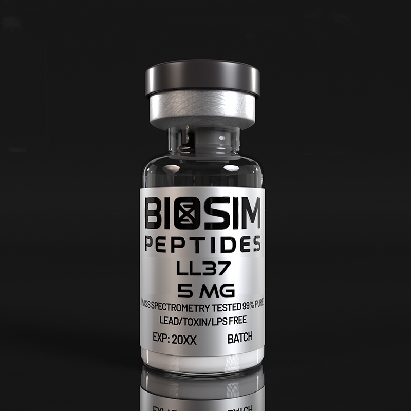
Research Overview
LL-37 is the sole human member of the cathelicidin family of antimicrobial peptides (AMPs) and represents one of the most extensively studied host defense peptides in innate immunity research. The peptide derives its name from its 37-amino-acid length and the presence of two N-terminal leucine residues (Leu-Leu). It is liberated from the C-terminal domain of its precursor protein, human cationic antimicrobial protein 18 (hCAP-18), through proteolytic processing by serine proteases including kallikrein 5 and proteinase 3 [1,5]. Unlike many antimicrobial peptides that function primarily through direct membrane disruption, LL-37 exhibits a remarkably broad functional repertoire encompassing direct antimicrobial activity against Gram-positive and Gram-negative bacteria, fungi, and enveloped viruses; potent immunomodulatory signaling through multiple receptors including Toll-like receptors (TLRs), formyl peptide receptor 2 (FPR2/ALX), and the P2X7 purinergic receptor; chemotactic recruitment of neutrophils, monocytes, and T cells; promotion of angiogenesis and re-epithelialization in wound healing; and disruption of pre-formed bacterial and fungal biofilms [1,3,4,5,6].
LL-37 expression is regulated by the vitamin D pathway through a vitamin D response element (VDRE) in the CAMP gene promoter, linking innate immune competence to vitamin D status — a regulatory mechanism uniquely prominent in primates [6]. This pleiotropic functionality positions LL-37 as a central integrator of the innate and adaptive immune responses, making it an indispensable research tool for investigators studying host-pathogen interactions, mucosal immunology, wound repair mechanisms, and immunomodulatory peptide biology.
Molecular Background & Mechanism of Action
The CAMP gene on chromosome 3p21.3 encodes the 170-amino-acid precursor protein hCAP-18, which comprises an N-terminal cathelin-like domain (highly conserved across species) and the C-terminal antimicrobial domain that, upon cleavage, yields the mature LL-37 peptide. In solution, LL-37 adopts a largely disordered conformation but undergoes a transition to an amphipathic α-helical structure upon interaction with lipid membranes or in membrane-mimetic environments, a structural rearrangement critical to its diverse biological activities [3]. The peptide carries a net positive charge of +6 at physiological pH, facilitating electrostatic interactions with negatively charged bacterial membrane components including lipopolysaccharide (LPS) in Gram-negative bacteria, lipoteichoic acid (LTA) in Gram-positive bacteria, and anionic phospholipids such as phosphatidylglycerol and cardiolipin.
The antimicrobial mechanism of LL-37 is multifaceted. At micromolar concentrations, the peptide inserts into bacterial membranes via a toroidal pore or “carpet” mechanism, disrupting the transmembrane electrochemical gradient and causing leakage of cytoplasmic contents [1,3]. Beyond direct membrane activity, LL-37 binds and neutralizes pro-inflammatory bacterial pathogen-associated molecular patterns (PAMPs) including LPS and LTA, effectively sequestering these TLR agonists and attenuating TLR4- and TLR2-mediated inflammatory signaling [4]. Simultaneously, LL-37 engages host cell receptors — most notably FPR2/ALX on neutrophils and monocytes, and the P2X7 receptor on macrophages and epithelial cells — to orchestrate chemotaxis, cytokine release, and inflammasome activation [4,5]. In the context of wound healing, LL-37 transactivates the epidermal growth factor receptor (EGFR) via metalloproteinase-mediated release of heparin-binding EGF-like growth factor (HB-EGF), promoting keratinocyte and epithelial cell migration and proliferation [2].
🔬 Mechanism at a Glance
- Gene: CAMP (cathelicidin antimicrobial peptide), chromosome 3p21.3 — regulated by vitamin D via VDRE in promoter
- Precursor: hCAP-18 (170 aa) → proteolytic cleavage by kallikrein 5 / proteinase 3 → mature LL-37 (37 aa)
- Structure: Disordered in aqueous solution → amphipathic α-helix upon membrane contact
- Direct Antimicrobial: Toroidal pore / carpet mechanism → membrane permeabilization at μM concentrations
- Immunomodulation: LPS/LTA neutralization (blocks TLR4/TLR2); FPR2/ALX activation (chemotaxis); P2X7 receptor engagement (inflammasome); EGFR transactivation (wound healing)
- Biofilm Activity: Disruption of pre-formed biofilms at sub-antimicrobial concentrations via matrix interaction and persister cell targeting
Preclinical & Clinical Evidence
The pleiotropic biology of LL-37 has been extensively characterized across a wide range of experimental systems. Fabisiak and colleagues (2016) provided a comprehensive review cataloguing LL-37’s activities spanning direct antimicrobial killing, LPS neutralization, chemotaxis, mast cell degranulation, angiogenesis, and wound repair, emphasizing its role as a multifunctional effector molecule at the interface of innate and adaptive immunity [1]. Shaykhiev and colleagues (2005) demonstrated that LL-37 stimulates airway epithelial cell proliferation and wound closure through EGFR-mediated signaling, establishing an early mechanistic framework for the peptide’s role in mucosal repair and barrier maintenance [2].
The anti-biofilm properties of LL-37 represent a research area of particular contemporary interest. Wang and colleagues (2014) elucidated the three-dimensional structural determinants underlying LL-37’s antibacterial and anti-biofilm activities, demonstrating that specific structural motifs within the peptide are differentially required for membrane disruption versus biofilm matrix interaction [3]. Scheenstra and colleagues (2020) published a definitive review of cathelicidin-mediated TLR modulation, detailing how these peptides fine-tune inflammatory responses through dual mechanisms: direct neutralization of TLR ligands (LPS, LTA, CpG DNA) and modulation of intracellular TLR signaling cascades including NF-κB and MAPK pathways [4]. Bowdish and colleagues (2005) provided an early, influential synthesis of LL-37’s role in anti-infective immunity, documenting its capacity to selectively modulate inflammatory gene expression — upregulating chemokines and anti-inflammatory cytokines while suppressing pro-inflammatory TNF-α responses to TLR agonists [5].
More recent investigations have expanded our understanding of LL-37 biology into translational contexts. Aloul and colleagues (2022) reviewed the evidence linking LL-37 expression to COVID-19 severity, highlighting the vitamin D–CAMP axis as a potential modulator of SARS-CoV-2-associated inflammatory responses, and demonstrating that LL-37 can bind the SARS-CoV-2 spike protein and inhibit viral entry in vitro [7]. The vitamin D-dependent regulation of CAMP gene expression, reviewed in multiple contexts including cancer biology by Chen and colleagues (2018), represents a unique evolutionary feature of the primate innate immune system linking nutritional status to antimicrobial defense capability [6].
Research Applications
LL-37 is supplied exclusively for in vitro and laboratory animal research. Investigators may employ this peptide in the following research domains:
- Antimicrobial Susceptibility Testing: Minimum inhibitory concentration (MIC) and minimum bactericidal concentration (MBC) determination against Gram-positive (e.g., S. aureus, MRSA), Gram-negative (e.g., P. aeruginosa, E. coli, A. baumannii), and fungal pathogens. Time-kill kinetic assays under varying ionic strength and pH conditions.
- Biofilm Research: Evaluation of LL-37-mediated dispersal and eradication of pre-formed mono- and polymicrobial biofilms using crystal violet staining, confocal microscopy with LIVE/DEAD staining, and colony-forming unit enumeration. Assessment of synergy between LL-37 and conventional antibiotics against biofilm-embedded persister cells.
- Innate Immunity & TLR Signaling: Investigation of LL-37-mediated LPS/LTA neutralization using TLR4/TLR2 reporter cell lines (e.g., HEK-Blue™ hTLR4/hTLR2). Quantification of NF-κB, MAPK, and IRF pathway modulation by Western blot, luciferase reporter, and cytokine multiplex assays.
- Wound Healing & Epithelial Biology: Scratch wound assays and electric cell-substrate impedance sensing (ECIS) for keratinocyte and epithelial cell migration. EGFR transactivation studies using phospho-EGFR ELISA and metalloproteinase inhibitor co-treatment paradigms.
- Vitamin D–Cathelicidin Axis: Investigation of 1,25(OH)₂D₃-mediated CAMP gene induction in epithelial, macrophage, and dendritic cell models using qRT-PCR, ChIP analysis of the CAMP promoter VDRE, and secreted LL-37 ELISA quantification.
- Immunomodulatory Peptide Pharmacology: Chemotaxis assays (Boyden chamber/Transwell) for neutrophil and monocyte migration. Flow cytometric analysis of leukocyte activation markers. Cytokine/chemokine profiling in LL-37-stimulated PBMCs and macrophage cell lines.
Comparative Context: LL-37 vs. Other Antimicrobial Peptides
Human Defensins (α-Defensins / HNP, β-Defensins / HBD)
Human defensins represent the other major family of antimicrobial peptides alongside cathelicidins. α-Defensins (HNP-1 through HNP-4, also known as human neutrophil peptides) are 29–35 residue, cysteine-rich, β-sheet peptides stored in neutrophil azurophilic granules, while β-defensins (HBD-1 through HBD-4) are expressed primarily by epithelial cells. Both families function through membrane disruption, but defensins are structurally constrained by three intramolecular disulfide bonds and generally exhibit narrower immunomodulatory profiles compared to LL-37. Parducho and colleagues (2020) demonstrated that HBD-2 inhibits Pseudomonas aeruginosa biofilm formation without compromising bacterial metabolic activity, revealing a mechanism distinct from the biofilm-disruptive and bactericidal action of LL-37 [8]. A key functional difference lies in LL-37’s capacity to neutralize LPS and modulate TLR signaling — properties less pronounced in the defensin family — making LL-37 a preferred research tool for studies at the intersection of antimicrobial activity and immunomodulation [1,4].
Histatins
Histatins are a family of histidine-rich, cationic peptides (7–38 amino acids) secreted by human salivary glands, with histatin-5 being the most potent antifungal member. Unlike LL-37, which targets a broad spectrum of bacteria, fungi, and viruses, histatins exhibit a narrower antimicrobial spectrum primarily focused on fungal pathogens, particularly Candida albicans. Histatins kill fungi through a non-lytic mechanism involving binding to fungal cell wall proteins, energy-dependent internalization, and targeting of mitochondrial ATP synthesis rather than membrane disruption. While both histatins and LL-37 contribute to oral mucosal immunity, LL-37’s broader receptor-mediated immunomodulatory activities — FPR2/ALX activation, TLR modulation, and EGFR transactivation — distinguish it from the more narrowly antifungal histatin family, positioning LL-37 as a superior model peptide for investigating the interface between innate immunity and tissue repair [1,5].
Safety & Handling Information
⚠️ FOR LABORATORY RESEARCH USE ONLY. This product is not approved for human use. Not for diagnostic or therapeutic purposes. Not for use in food-producing animals.
- Appearance: White to off-white lyophilized powder
- Purity: ≥95% as determined by RP-HPLC
- Sequence: LLGDFFRKSKEKIGKEFKRIVQRIKDFLRNLVPRTES (37 amino acids; single-letter code)
- Molecular Formula: C₂₀₅H₃₄₀N₆₀O₅₃
- Molecular Weight: 4,493.3 Da
- Net Charge: +6 at physiological pH (~7.4)
- Storage: Lyophilized peptide should be stored at -20°C, protected from light and moisture. LL-37 is hygroscopic; ensure container is tightly sealed after each use. Reconstituted solutions should be aliquoted, stored at -20°C to -80°C, and subjected to minimal freeze-thaw cycles.
- Reconstitution: LL-37 is freely soluble in sterile water. For cell-based assays, stock solutions may be prepared in sterile water or 0.01% acetic acid and subsequently diluted into appropriate cell culture medium. The peptide may adsorb to plastic surfaces; use of low-protein-binding tubes and tips or inclusion of carrier protein (e.g., 0.1% BSA) in working solutions is recommended for experiments requiring precise concentration control.
- Stability: Lyophilized peptide is stable for 12–24 months when stored as recommended. LL-37 is susceptible to proteolytic degradation in complex biological matrices; researchers should validate peptide stability under their specific experimental conditions and consider the use of protease inhibitors where appropriate.
- Handling Precautions: Use appropriate personal protective equipment (PPE) including gloves, lab coat, and eye protection. Handle in a biosafety cabinet or fume hood. Avoid inhalation of powder and contact with skin or eyes. As a bioactive immunomodulatory peptide, avoid accidental exposure.
References
- PMID: 27117377 — Fabisiak A, Murawska N, Fichna J. LL-37: Cathelicidin-related antimicrobial peptide with pleiotropic activity. Pharmacol Rep. 2016;68(4):802-808. doi:10.1016/j.pharep.2016.03.015
- PMID: 15964896 — Shaykhiev R, Beisswenger C, Kändler K, et al. Human endogenous antibiotic LL-37 stimulates airway epithelial cell proliferation and wound closure. Am J Physiol Lung Cell Mol Physiol. 2005;289(5):L842-L848. doi:10.1152/ajplung.00186.2005
- PMID: 24463069 — Wang G, Mishra B, Epand RF, et al. High-quality 3D structures shine light on antibacterial, anti-biofilm and antiviral activities of human cathelicidin LL-37 and its fragments. Biochim Biophys Acta. 2014;1838(9):2160-2172. doi:10.1016/j.bbamem.2014.01.016
- PMID: 32582207 — Scheenstra MR, van Harten RM, Veldhuizen EJA, et al. Cathelicidins Modulate TLR-Activation and Inflammation. Front Immunol. 2020;11:1137. doi:10.3389/fimmu.2020.01137
- PMID: 15569695 — Bowdish DM, Davidson DJ, Lau YE, et al. Impact of LL-37 on anti-infective immunity. J Leukoc Biol. 2005;77(4):451-459. doi:10.1189/jlb.0704380
- PMID: 29843147 — Chen X, Zou X, Qi G, et al. Roles and Mechanisms of Human Cathelicidin LL-37 in Cancer. Cell Physiol Biochem. 2018;47(3):1060-1073. doi:10.1159/000490183
- PMID: 35634307 — Aloul KM, Nielsen JE, Defensor EB, et al. Upregulating Human Cathelicidin Antimicrobial Peptide LL-37 Expression May Prevent Severe COVID-19 Inflammatory Responses and Reduce Microthrombosis. Front Immunol. 2022;13:880961. doi:10.3389/fimmu.2022.880961
- PMID: 32457749 — Parducho KR, Beadell B, Ybarra TK, et al. The Antimicrobial Peptide Human Beta-Defensin 2 Inhibits Biofilm Production of Pseudomonas aeruginosa Without Compromising Metabolic Activity. Front Immunol. 2020;11:951. doi:10.3389/fimmu.2020.00951
⚠️ Research Use Only — Important Disclaimer
This product is intended solely for laboratory research purposes and is not manufactured, tested, or certified for human or veterinary use. It is not a drug, food, dietary supplement, or cosmetic. Researchers must comply with all applicable institutional, local, state, and federal regulations governing the acquisition, storage, handling, and disposal of research peptides. Biosim Peptides makes no representations regarding the suitability of this product for any purpose other than bona fide scientific research conducted by qualified professionals in appropriate laboratory facilities. No statements on this page have been evaluated by the FDA or any other regulatory agency.
Frequently Asked Questions
Q: What is the relationship between LL-37 and hCAP-18?
hCAP-18 (human cationic antimicrobial protein, 18 kDa) is the 170-amino-acid precursor protein encoded by the CAMP gene. LL-37 is the 37-amino-acid C-terminal antimicrobial domain liberated from hCAP-18 through proteolytic cleavage by serine proteases including kallikrein 5 (in skin) and proteinase 3 (in neutrophils). hCAP-18 itself is stored in neutrophil secondary granules and secreted by epithelial cells; the mature LL-37 peptide is the biologically active effector molecule [1,5].
Q: How does vitamin D regulate LL-37 expression?
The CAMP gene promoter contains a vitamin D response element (VDRE) that binds the vitamin D receptor (VDR)–retinoid X receptor (RXR) heterodimer upon activation by 1,25-dihydroxyvitamin D₃ (calcitriol). This transcriptional regulatory mechanism directly links vitamin D status to LL-37 production in epithelial cells, macrophages, and other innate immune effector cells. This VDRE-mediated regulation is a primate-specific evolutionary feature not found in rodents, which is an important consideration when selecting experimental models [6].
Q: What concentration of LL-37 is appropriate for antimicrobial assays?
Reported MIC values for LL-37 vary considerably depending on bacterial strain, inoculum size, growth medium composition, and ionic strength. Typical active concentrations range from 1–64 μM against susceptible Gram-positive and Gram-negative organisms in low-ionic-strength buffers, with higher concentrations required in physiological salt conditions (150 mM NaCl) or complex media. Researchers should perform pilot dose-ranging experiments to establish appropriate working concentrations for their specific pathogen–medium combinations [1,3].
Q: How does LL-37 compare to conventional antibiotics for biofilm research?
Unlike many conventional antibiotics that target metabolically active cells in specific growth phases, LL-37 can disrupt pre-formed biofilms through multiple mechanisms: interaction with the extracellular polymeric substance (EPS) matrix, permeabilization of biofilm-embedded cells including metabolically dormant persisters, and immunomodulatory signaling that potentiates host-mediated clearance. These properties make LL-37 a valuable research tool for studying biofilm biology and evaluating anti-biofilm strategies that complement or synergize with conventional antimicrobial agents [3].
Q: Does LL-37 require specific handling to prevent adsorption to labware?
Yes. LL-37, like many cationic amphipathic peptides, can adsorb nonspecifically to plastic and glass surfaces, potentially reducing effective concentrations in experimental systems. Recommended mitigation strategies include: use of low-protein-binding (siliconized or LoBind) microcentrifuge tubes and pipette tips; inclusion of 0.01–0.1% bovine serum albumin as a carrier in working solutions; and preparation of fresh working dilutions immediately before use rather than extended storage of dilute solutions. Researchers should validate peptide recovery in their specific experimental formats using appropriate analytical methods (RP-HPLC or ELISA).

Reviews
There are no reviews yet.