Research Overview
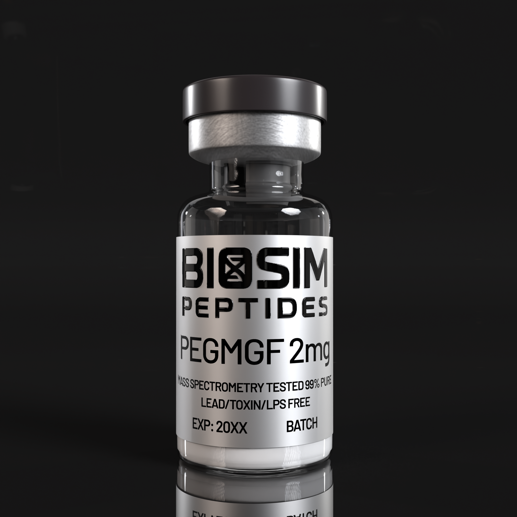
PEG-MGF (Pegylated Mechano Growth Factor) represents a significant advancement in the study of muscle repair and regenerative biology. Mechano Growth Factor (MGF) is the alternative splice variant of the insulin-like growth factor 1 (IGF-1) gene, specifically the IGF-1 Ec isoform in humans (rodent homolog: IGF-1 Eb). Unlike systemic, liver-derived IGF-1, MGF is expressed locally in skeletal muscle in direct response to mechanical stretch and tissue microdamage, making it a focal point of research into muscle satellite (stem) cell activation, myogenesis, and tissue restoration. The PEGylation — covalent attachment of polyethylene glycol chains — extends the peptide’s biological half-life by reducing renal clearance and proteolytic degradation, thereby enabling sustained signaling in experimental models. PEG-MGF 2mg is supplied as a lyophilized powder for laboratory research use only, not for human consumption or clinical application.
Molecular Background & Mechanism of Action
The IGF-1 gene undergoes tissue-specific alternative splicing to produce distinct mRNA transcripts. In the liver, the predominant transcript yields circulating IGF-1Ea, responsible for systemic endocrine growth signaling. In skeletal muscle subjected to mechanical overload, stretching, or injury, a splice shift produces the IGF-1 Ec transcript — encoding MGF — which includes a unique C-terminal E-peptide extension not found in the systemic isoform. This C-terminal domain is functionally autonomous and mediates MGF’s distinct biological activities independent of IGF-1 receptor signaling (Matheny et al., 2010).
Following muscle microtrauma, MGF is rapidly upregulated within hours, preceding the later rise in IGF-1Ea. This temporal pattern suggests a two-phase repair model: an early MGF-driven satellite cell activation and proliferation phase, followed by an IGF-1Ea-mediated differentiation and hypertrophy phase. PEGylation of the MGF peptide obscures sites vulnerable to circulating proteases and increases hydrodynamic radius to reduce glomerular filtration, yielding a sustained signaling window that researchers can leverage to study extended satellite cell activation kinetics and myogenic progression in longer-duration experimental protocols.
MGF binds to and activates muscle satellite (stem) cells via signaling pathways distinct from classical IGF-1 receptor cascades. The unique C-terminal E-peptide domain stimulates satellite cell proliferation and migration to injury sites, while also exerting neuroprotective effects in neuronal injury models. PEGylation prolongs the peptide’s circulating half-life from minutes to hours, permitting sustained receptor engagement. In contrast to IGF-1 LR3 — a modified IGF-1 with high systemic IGF-1 receptor affinity — MGF operates through an autocrine/paracrine mechanism localized to mechanically stressed tissues, and regular (non-pegylated) MGF exhibits a shorter window of activity. This makes PEG-MGF a uniquely suited tool for investigating prolonged muscle repair signaling.
Preclinical & Experimental Evidence
The foundational work by Hill and Goldspink (2003) established that MGF expression and IGF-1 gene splicing are directly associated with satellite cell activation following local tissue damage in rodent skeletal muscle. Their immunohistochemical analyses demonstrated that MGF peptide is the first IGF-1 isoform produced at damage sites, with peak expression occurring within 24 hours of injury and co-localizing with activated satellite cell markers.
In human studies, Hameed et al. (2003) profiled IGF-I splice variant expression in young and old human skeletal muscle after high-resistance exercise, finding that MGF upregulation after mechanical loading is significantly attenuated in aged muscle — a finding with direct implications for sarcopenia research. This age-associated decline in MGF responsiveness was further detailed by Goldspink (2006), who characterized the impairment of IGF-I gene splicing mechanisms in muscle wasting conditions, establishing MGF deficiency as a molecular correlate of regenerative failure.
Kandalla et al. (2011) demonstrated that the MGF E-peptide directly activates human muscle progenitor cells regardless of donor age, increasing both proliferative capacity and myogenic fusion potential. This suggests that exogenous MGF administration may bypass age-related deficiencies in endogenous MGF production, a hypothesis now testable with PEG-MGF in controlled laboratory settings.
Beyond striated muscle, MGF has demonstrated notable neuroprotective properties. Dluzniewska et al. (2005) showed that the autonomous C-terminal peptide of IGF-1 Ec exerts potent neuroprotection in experimental brain ischemia. In a motor neuron disease context, Riddoch-Contreras et al. (2009) found that MGF administration rescued motoneurons and improved muscle function in the SOD1(G93A) mouse model of amyotrophic lateral sclerosis (ALS), while Ates et al. (2007) demonstrated that MGF treatment increased progenitor cell populations in both dystrophic and ALS-affected muscle, as well as in normal muscle tissue — underscoring its broad regenerative potential.
Matheny et al. (2010) provided an authoritative minireview consolidating the evidence for MGF as a tissue repair and regeneration factor, delineating its roles in muscle, bone, nerve, and cardiac tissue, and noting that the unique E-peptide domain constitutes a compelling target for therapeutic peptide engineering — of which PEGylation is a primary strategy.
Comparison: PEG-MGF vs. IGF-1 LR3 vs. Regular MGF
While IGF-1 LR3 is an engineered IGF-1 analog with an arginine substitution at position 3 and an N-terminal 13-amino-acid extension that reduces binding to IGF binding proteins (IGFBPs) and increases systemic bioavailability, it signals primarily through the classical IGF-1 receptor and activates the PI3K/Akt pathway systemically. In contrast, MGF’s unique E-peptide mediates receptor interactions that are at least partially independent of the IGF-1 receptor, driving satellite cell activation rather than bulk myofiber hypertrophy. Regular (non-pegylated) MGF has a very short in vivo half-life measured in minutes due to rapid proteolytic cleavage of the C-terminal E-peptide, limiting its utility in extended-duration experiments. PEG-MGF addresses this limitation through polyethylene glycol conjugation, which shields the E-peptide domain from enzymatic degradation and extends the research window considerably, making it the preferred format for studies requiring sustained MGF signaling over hours to days.
Research Applications
- Muscle Satellite Cell Biology: Investigate the kinetics of satellite cell activation, proliferation, and myogenic lineage commitment under sustained MGF signaling conditions.
- Sarcopenia & Age-Related Muscle Wasting: Study whether exogenous PEG-MGF can compensate for age-related declines in endogenous MGF expression and restore regenerative capacity in aged muscle tissue models.
- Muscular Dystrophy Research: Examine the effects of prolonged MGF signaling on muscle progenitor cell populations in dystrophic muscle models, including Duchenne and Becker muscular dystrophies.
- Motor Neuron & Neuromuscular Research: Explore the neuroprotective and motoneuron-rescuing properties of the MGF E-peptide in models of ALS, spinal muscular atrophy, and peripheral nerve injury.
- Cardiac Muscle Repair: Investigate MGF’s role in cardiac myocyte protection and regeneration following ischemia-reperfusion injury in cardiac tissue models.
- Bone & Tendon Regeneration: Evaluate the effects of sustained MGF delivery on osteoblast proliferation and tenocyte repair in musculoskeletal injury models.
- PEGylation Technology Studies: Use PEG-MGF as a model peptide to study the effects of PEGylation on peptide stability, tissue distribution, and sustained-release kinetics in comparative pharmacology experiments.
Safety & Laboratory Handling
PEG-MGF is supplied as a sterile, lyophilized powder in a sealed glass vial. Standard laboratory safety protocols should be observed, including the use of appropriate personal protective equipment (PPE): laboratory coat, nitrile gloves, and safety goggles. The lyophilized powder should be stored at -20°C for long-term stability, protected from light and moisture. Upon reconstitution with an appropriate solvent (e.g., sterile bacteriostatic water or buffer solution), PEG-MGF solution should be kept refrigerated at 2–8°C and used within the timeframe established by the researcher’s stability protocol. Avoid repeated freeze-thaw cycles, which may compromise peptide integrity. Handle in a biosafety cabinet or laminar flow hood to maintain sterility. Dispose of all materials in accordance with institutional guidelines for laboratory reagents.
References
- Matheny RW Jr, Nindl BC, Adamo ML. Minireview: Mechano-growth factor: a putative product of IGF-I gene expression involved in tissue repair and regeneration. Endocrinology. 2010 Mar;151(3):865-875. PMID: 20130113.
- Hill M, Goldspink G. Expression and splicing of the insulin-like growth factor gene in rodent muscle is associated with muscle satellite (stem) cell activation following local tissue damage. J Physiol. 2003 Jun 1;549(Pt 2):409-418. PMID: 12692175.
- Hameed M, Orrell RW, Cobbold M, Goldspink G, Harridge SD. Expression of IGF-I splice variants in young and old human skeletal muscle after high resistance exercise. J Physiol. 2003 Feb 15;547(Pt 1):247-254. PMID: 12562960.
- Ates K, Yang SY, Orrell RW, Sinanan AC, Simons P, et al. The IGF-I splice variant MGF increases progenitor cells in ALS, dystrophic, and normal muscle. FEBS Lett. 2007 Jun 12;581(14):2727-2732. PMID: 17531227.
- Dluzniewska J, Sarnowska A, Beresewicz M, Johnson I, Srai SK, et al. A strong neuroprotective effect of the autonomous C-terminal peptide of IGF-1 Ec (MGF) in brain ischemia. FASEB J. 2005 Nov;19(13):1896-1898. PMID: 16144956.
- Riddoch-Contreras J, Yang SY, Dick JR, Goldspink G, Orrell RW, et al. Mechano-growth factor, an IGF-I splice variant, rescues motoneurons and improves muscle function in SOD1(G93A) mice. Exp Neurol. 2009 Feb;215(2):281-289. PMID: 19038252.
- Kandalla PK, Goldspink G, Butler-Browne G, Mouly V. Mechano Growth Factor E peptide (MGF-E), derived from an isoform of IGF-1, activates human muscle progenitor cells and induces an increase in their fusion potential at different ages. Mech Ageing Dev. 2011 Apr;132(4):154-162. PMID: 21354439.
- Goldspink G. Impairment of IGF-I gene splicing and MGF expression associated with muscle wasting. Int J Biochem Cell Biol. 2006 Mar;38(3):481-489. PMID: 16463438.
PEG-MGF 2mg is intended exclusively for in vitro laboratory research. This product is not a drug, food supplement, or cosmetic. It is not for human or veterinary diagnostic, therapeutic, or prophylactic use. All handling must be conducted by qualified laboratory personnel in accordance with institutional biosafety and chemical hygiene protocols. Biosim Peptides does not endorse or encourage human consumption under any circumstances.

Reviews
There are no reviews yet.